
Liver dysfunction in MZs
Pericentral Dysfunction and Systemic Consequences in Pi*MZ

News & Research Update
2 August, 2025
Dear readers and MZ patients, last week we promised to come out with a more detailed and simplified explanation about:
“Understanding Pi*MZ Liver Dysfunction: A UK Biobank–Supported Model of Functional Impairment”
of which you received the overview last weekend.
So, we are now starting explaining the details in a series of newsletters of the coming weeks. We suggest you keep them stored for later as well…
Recruitment-Secretory Block in the Liver (root cause MZ)
Functional Capacity of the Liver (aging, pregnancy, medication, and illness/inflammation)
Functional Cholestasis
Impaired Calcium Absorption and Vitamin D deficiency
Fat-Soluble Vitamin Deficiencies
Small Intestinal Bacteria Overgrowth (SIBO)
Vitamin B12 deficiency
Impaired red blood cell production & oxygen transport
Thrombocyte production
Hormonal imbalance
Food and lifestyle recommendations
Week 1: The Recruitment Secretory Block: The Root Cause in Pi*MZ
1. Your Liver’s Working Units
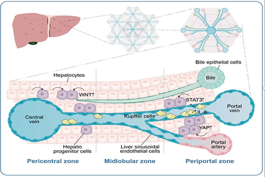
Let's first understand some basics of your liver. Your liver is made up of tiny building blocks called lobules. Each lobule has three zones with liver cells called hepatocytes, and each zone has its typical job:
· The periportal zone (zone 1) produces many essential proteins, including alpha‑1 antitrypsin (AAT).
· The midlobular zone (zone 2) is more of a reserve area that helps when demand is high, and plays a role in liver regeneration.
· The pericentral zone (zone 3) handles detoxification, hormone processing, and bile acid production.
Below is a picture of the liver, divided into segments, and finally at the bottom, a single element, the liver lobule, with the liver cells called hepatocytes.
2. What Happens in an MZ Liver
If you are an Alpha 1 MZ, your liver cells (hepatocytes) produce two types of AAT:
Normal M‑AAT, which is secreted into your blood.
Misfolded Z‑AAT, which gets trapped inside liver cells.
Your cells try to clean up this misfolded protein through a process called autophagy (a cellular recycling system). Some people do this more effectively than others. The faster the clean‑up, the less stress on the liver, which explains why some MZs have more issues than others. Current indications are that about 25% of the MZ population has liver problems.
3. The Recruitment-Secretory Block
When your body is under stress, for example because of inflammation or infection, the liver will produce more AAT.
At first, zone 1 cells will recruit more liver cells to increase production.
Soon after, liver cells in zones 2 and 3 are also recruited to help.
But here’s the problem:
These newly recruited cells (hepatocytes) also produce Z‑AAT.
Therefore, the misfolded protein accumulates in them as well.
The stress spreads from the periportal (zone 1) into the midlobular zone (zone 2) and into the pericentral zone (zone 3).
This is called the Recruitment-Secretory Block (R-SB). Instead of helping, the recruited cells become also burdened, reducing the liver’s overall capacity and increasing the stress in the liver (which is also an inflammatory process).
4. Why It Matters
Zone 3 is more responsible for detoxifying medicines, regulating hormones, and regulating bile acid, while zone 2 is more responsible for regenerating the liver after injury. When they are busy producing AAT and handling the Z‑AAT stress, the liver’s ability to do these other essential tasks is reduced. Over time, this can contribute to health issues, even if your standard liver tests look “normal.”
5. What Research Shows
Callea et al. (2021) demonstrated under the microscope that Z‑AAT inclusions appear not only in zone 1, but also in the midlobular and pericentral zones. [1]
Sanders et al. (2019) showed that during inflammation, AAT levels rise in MZ individuals but not in ZZ. This rise is proof that more liver cells are being recruited. [2]
This extra recruitment is the key driver of whole‑lobule stress in Pi*MZ.
Note: In MZ, higher blood AAT levels may sound reassuring, but they can actually be a warning sign, showing that the liver is working harder and more liver cells will be under stress. We’ll explain this in more detail in a later newsletter.
The image from the study of Callea et al. (figure 1) shows a cross-section of a very small part of the liver, with the periportal part (zone 1) of the liver lobule on the right and the pericentral part (zone 3) of the liver lobule on the left. The black dots clearly show the AAT inclusions affecting the whole liver lobule from right to left.
The image from the study of Sanders et al. (2019) (figure 2) shows the level of AAT in the blood depending on the level of inflammation.
The lower line (purple) shows clearly that the Alpha-1 ZZ liver is not really capable of increasing the AAT much, and the amount of AAT is even decreasing when the inflammation worsens (CRP is a measure of inflammation).
Instead, the red line (MZ) shows an increase in AAT with inflammation, indicating the recruitment of new liver cells.
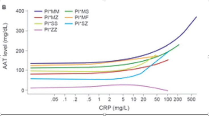
Figure 2 Sanders et al, 2019
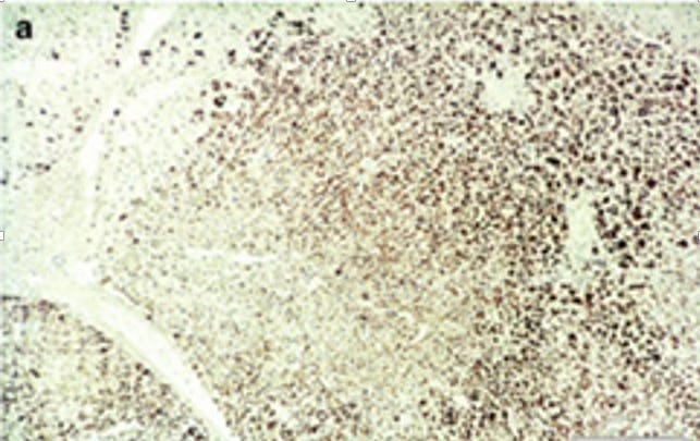
Figure 3 Int J Mol Sci. 2021 Jun 24;22(13):6807. doi: 10.3390/ijms22136807
6. Key Takeaway
Pi*MZ is not a neutral state. The Recruitment-Secretory Block shows that hepatic stress spreads across the entire liver lobule, reducing the liver's capacity for other tasks. This matches the morbidities seen in the UK Biobank. Recognizing and understanding this key element is the first and foremost important step in understanding the MZ patient.
Did you find this article interesting? Then please don’t forget to subscribe to our newsletter and share it with others! In our next newsletter, we will tell you all about the functional capacity of the liver, an interesting topic for all Alpha’s.
Since MZs can also have lung issues, we will definitely pay attention to that part of alpha-1 antitrypsin deficiency in our newsletters. To be continued…
And, like always, enjoy the ride!!
[1] Callea et al. (2021) – The Recruitment-Secretory Block (“R-SB”) Phenomenon and Endoplasmic Reticulum Storage Diseases
DOI: 10.3390/ijms22136807
[2] Sanders et al. (2019) – The Effects of Inflammation on Alpha 1 Antitrypsin Levels in a National Screening Cohort
DOI: 10.1080/15412555.2017